|
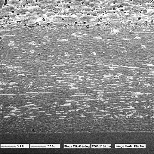
This FIB secondary electron image shows a FIB cross-section of a sample of colour photographic film. No special
specimen preparation was required; a piece of film was inserted into the FIB, and sectioning took less than ten
minutes. The FIB stage was then tilted, in-situ, to 45° to allow viewing of the section surface. A porous surface
layer, and layers of "colour absorbing grains," lying primarily in the plane of the film, are clearly distinguishable.
As this sample demonstrates, FIB specimen preparation and imaging is not limited to conductive, metallic samples.
Using charge neutralization when necessary, non-conductive ceramic, polymeric and geological specimens may be
examined. Success has also been reported in the literature on the use of cold stages to example "wet" biological
materials.
|