|
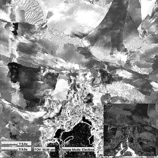
Comparative FIB images of a crack in a pipeline steel, acquired using secondary electron and secondary ion signals.
The ion image (inset) highlights the corrosion product in the crack
itself, while the electron image shows the microstructural features within the plastic zone around the crack tip. This
sample was metallographically polished prior to FIB imaging.
FIB secondary ion images are particularly sensitive to the presence of oxides and carbides in metallic systems due to
the effect these elements have on the secondary ion yield of the metal. The oxygen or carbon "enhanced yield" can
cause order-of-magnitude or more increases in the brightness of a region containing metal oxides or carbides,
creating a chemical contrast effect that makes FIB SII an ideal technique for identifying corrosion or grain boundary
segregation without resorting to chemical etching. Eliminating the need to use chemical etching is significant as many
etchants contain elements that are also expected to be
found in the native corrosion products. Etching frequently contaminates the sample to a depth of several micrometers
into precipitates or porous grain boundaries. FIB SII and SEI imaging combined with careful metallographic
polishing techniques that minimize mechanical deformation introduced during preparation prior to FIB work permits
the use of FIB micromachining to produce TEM specimens with virtually a one-to-one correspondence between the
TEM specimen and the FIB imaged region - without concern that the chemical analysis to be performed in the TEM
will be compromised by chemicals in a conventional etchant.
|