Fibics Pattern
TEM Image of a Microprocessor
Prepared by FIB
This is a transmission electron microscope image of a modern microprocessor. The cross-section was prepared using a FIB to precisely section the transistor at the bottom left corner. Specimens like this site specific TEM sample can be prepared to only 25 nano-metres thick.
TEM Image of a Microprocessor Prepared by FIB
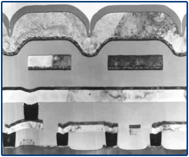
This image demonstrates the two real advantages of fabricating samples using a FIB microscope. A reasonable amount of control over the final thickness can be exercised by using the FIB, as well as the great advantage of site-specificity, the location of the TEM sample can be selected with sub-micron positional accuracy.